One of the earliest uses of Cord Lining Stem Cells is in resurfacing burns. The Stem Cells are placed on a flexible scaffold and then applied onto the wound after surgery. This enhances recovery of burns without requiring a skin graft from another part of the body. Stem cell treatment is commonly used for burn victims with chronic or widespread burns.


Examples of successful treatments of burns using our stem cell technology:

  1. 53 year old lady with chronic thermal burn wound was treated with Cord Lining Stem Cells. The wound was closed in seven days, no skin graft was required, and the wound has continued to remain stable to this day - almost five years later.
  2. 30 year old male with full thickness chemical burn wound on the dorsum of the foot, which had remained open for several months. The wound was successfully closed in 21 days with CLSC, and no skin graft was required. The patient was spared more complicated surgery (e.g. microvascular free flap resurfacing).


CellResearch is actively conducting research on burns applications using Cord Lining Stem Cells with these clinical collaborators:

  1. Professor David Herndon and Dr Katsuhiro Kita at the Shriners Hospital for Children in Galveston, Texas;
  2. Dr. Mai Manh Tuan at St Paul’s Hospital Burns Centre and the National Hospital of Traditional Medicine in Hanoi, Vietnam;

  3. Dr. Dinh Van Han at Hospital 103 in Hanoi, Vietnam;

  4. Professor Andrew Burd and Dr. Linda Huang at the Chinese University of Hong Kong in Hong Kong, SAR.


Our initial success with burns strongly suggested that the Cord Lining Stem Cells could also resurface chronic wounds and ulcers. The examples below show successful treatments we have achieved on ulcers and wounds where conventional treatments had failed. We hypothesised that the Cord Lining Stem Cells were secreting proteins that had modulated the chronic wound environment, making it amenable for repair.


Examples of successful treatments of wounds using our stem cell technology:


  1. 2 year old male with a chronic ulcer on the ankle after being treated for a blood vessel tumour, called a vascular malformation, with external irradiation. The vascular malformation shrank but a gaping wound was left in its wake. Treatment with Cord Lining Stem Cells closed the wound in 20 days after it had failed to heal for 3 months.
  2. 14-year-old male with a clotting disorder called Haemophilia. Patients with this condition are unable to clot their blood and can die from uncontrolled bleeding. In his history he bumped his ankle, which accumulated a blood clot (called a haematoma). This broke down revealing a large ulcer. His surgeons were unable to take a skin graft for fear he might bleed uncontrollably from where the skin graft was harvested (called the skin graft donor site). Treatment with Cord Lining Stem Cells healed the wound in 42 days without need for a skin graft.

  3. 30-year-old female who sustained severe injuries in a road traffic accident when she was flung from a motorcycle, sustaining arm fractures and tissue loss. Bone was exposed and skin grafting on bone was not feasible so her surgeons suggested amputation. Fearful of losing her forearm, this young lady opted to use Cord Lining Stem Cells, which successfully closed the wound over the exposed bone after 21 days, saving her from a forearm amputation.


CellResearch is actively conducting chronic wound resurfacing applications using Cord Lining Stem Cells with these clinical collaborators:

  1. Dr. Mai Manh Tuan at St Paul’s Hospital Burns Centre and the National Hospital of Traditional Medicine in Hanoi, Vietnam;
  2. Dr. Dinh Van Han at Hospital 103 in Hanoi, Vietnam


It has been said that ageing is paying for a crime you did not commit! Our joints are lined with cartilage, and bear the mechanical stresses as we move or run around from day to day. Cartilage takes a long time to heal, and if a small injury is inflicted, the cartilage may be progressively worn down before it has a chance to adequately repair itself. Ultimately, the underlying bone becomes damaged, and the joint undergoes degenerative arthritis in a condition called Osteoarthritis. Arthritis can also occur in inflammatory conditions like Rheumatoid Arthritis.


Cord Lining Stem Cells have been successfully differentiated into bone cells (osteocytes) and cartilage cells (chondrocytes). It is our hope that one day these cells may be directly transplanted to affected areas to replace the damaged bone and cartilage in arthritic conditions.


Our collaborators Dr Martin J. Stoddart and Professor Mauro Alini at the AO Research Institute in Davos, Switzerland are working to fine-hone Cord Lining Stem Cell differentiation into osteocytes and chondrocytes, and Professor James Richardson at Keele University in Staffordshire, England is researching clinical transplant applications for cartilage repair.  The FBM Regenerative Biology and Medicine JSC in Hanoi, Vietnam is also researching Cord Lining Stem Cells for cartilage repair and replacement.

Future applications include the integration of Cord Lining Stem Cells into synthetic bone grafts (used to fill bone defects after loss from tumour or trauma) to optimise synthetic bone graft take. This will reduce or even remove the need for painful bone graft harvest from other parts of the body.


We have collaborated with the Singapore National Eye Centre (SNEC) and Vietnam Eye Institute to develop applications where stem cell regeneration can be used to aid in human cornea repair. Cornea is the transparent front of the eye where light enters to fall onto the retina. Scarring or fogging of the cornea can result in diminished eyesight or even blindness. Cornea-related disease and degeneration increases with age. Alternative treatment methods using autologous stem cells reduce the need for corneal transplants.

Two papers have been published related to the use of Cord Lining Stem Cells in corneal resurfacing.

The first paper comprehensively characterised Cord Lining Stem Cells to show their stem cell features multipotent nature.

Publication: Reza HM, Ng B-Y, Phan TT, et al. Characterization of a novel umbilical cord lining cell with CD227 positivity and unique pattern of P63 expression and function. Stem Cell Rev. 2011 Sep; 7(3): 624-638

The second paper describes the successful transplantation of Cord Lining Stem Cells on amniotic membrane to resurface rabbit corneal defects.

Publication: Reza HM, Ng B-Y, Gimeno FL, et al. Umbilical cord lining stem cells as a novel and promising source for ocular surface regeneration. Stem Cell Rev. 2011 Nov; 7(4): 935-947

In Associate Professor Ang’s of the Singapore Eye Research Institute (SERI) of the Singapore National Eye Centre words:

“Our study introduces a novel umbilical cord derived cell with high proliferative capacity which may have potential therapeutic applications for use in regenerative medicine and tissue replacement for treating various diseases.”

This work was subsequently translated for human cornea repair performed at the Vietnam National Eye Institute by Associate Professor Hoang Minh Chau in association with the FBM Regenerative Biology and Medicine JSC, which is ongoing to date. So far, results have been excellent.

Associate Professor Chau and her team presented their data in their paper “Using Tissue- cultured Cord Lining Epithelial Cells in the Treatment of Persistent Corneal Epithelial Defect” on the resurfacing of 16 human eyes, 15 of which were treated successfully, at the Asia Cornea Society Meeting in the Philippines.


Diabetes Mellitus is a disease that afflicts more than 171 million people in the world, It causes high sugar levels in the blood, and this in turn causes problems with blood flow in small vessels (microvascular disease) which result in kidney, heart and nerve problems, as well as large ones (macrovascular disease) resulting in blood vessel blockage causing strokes, heart attacks and leg ulcers. Diabetes also, weakens the immune system, making patients susceptible to infections and prone to poor wound healing. 

Insulin is the hormone lacking in Diabetes patients, and they need to replace insulin by injecting themselves regularly with insulin. Insulin allows the cells of the body to absorb glucose from the blood for normal cellular activity.

Cord Lining Stem Cells have been successfully differentiated into Islet Cells that produce insulin. These differentiated islet cells can potentially be transplanted into patients with diabetes to produce insulin within the body without the need for regular injections.

Alternatively, the insulin gene may be directly inserted into Cord Lining Stem Cells to stimulate the cells to produce insulin before they are transplanted into the body using the process of transfection.

Currently, CellResearch Corporation is working with Professor Sir Roy Calne and Professor KO Lee at the National University of Singapore, as well as Professor Kon Oi Lian at the National Cancer Centre, Singapore looking specifically into this exciting area. In the not-too-distant future, Cord Lining Stem Cells could be the solution to this difficult disease.

These data have now also been published in a paper that shows that Cord Lining Epithelial Cells, in addition to being successfully differentiated into pancreatic islet cells, also produce immunosuppressant HLA called HLA-G and HLA-E which prevent rejection of transplanted Cord Lining Epithelial Cells from the recipient of these cells.

Publication: Zhou Y, Gan SU, Lin G, et al. Characterization of human umbilical cord- lining derived epithelial cells and transplantation potential. Cell Transplantation. 2011; 20: 1827-1841

To take a quote from the publication:

“In conclusion, the combination of the in vitro and in vivo data presented, plus the ease of CLEC derivation, demonstrates the potential of CLECs as a new candidate for cell transplantation.”


Haemophilia A is a blood clotting disease due to a genetic lack of production of Factor VIII, a crucial blood-clotting factor. This is called a X-linked recessive disease and the large majority of sufferers are male.


There is no cure for Haemophilia A, and haemophiliacs require regular, expensive transfusions of Factor VIII to survive, failing which even the slightest injury to the body can lead to uncontrolled or even fatal bleeding. Sometimes, antibodies are generated against replacement Factor VIII, so even more complex and expensive therapies are required to overcome this problem. Transfusion itself of contaminated blood has also resulted in transmission of the dreaded HIV (AIDS) virus.

Professor Kon Oi Lian at the National Cancer Centre has successfully implanted the Factor VIII gene into Cord Lining Stem Cells, and transplantation of these cells into mice have maintained levels of Factor VIII for up to 14 days.

This is the first step in research, with the ultimate aim of transplanting these cells into man to provide a long-term replacement of Factor VIII for sufferers of Haemophilia A.

The above work by Professor Kon has been published and not only shows successful integration into Cord Lining Epithelial Cells of the Factor VIII gene into haemophiliac mice and improvement of their clotting characteristics, but also shows the robustness of these cells as inserting the Factor VIII gene would damage weaker cells.

Publication: Sivalingam J, Krishnan S, Ng WH, et al. Biosafety assessment of site-directed transgene integration in human umbilical cord-lining cells. Mol Ther. 2010; (18)7: 1346-1356

This study will be followed by a dog study, after which human trials will follow.


The heart is the most emotive of organs in the human body. Its daily pumping function keeps us alive, and this is certainly a pump which never rests. By nature of its constant activity, anything that disrupts blood supply to the heart muscle can cause the heart muscle to suffocate from lack of oxygen (called myocardial ischaemia). When the muscles die in just one spot, they are replaced by weak fibrous tissue. Where large areas of the heart muscle dies, the patient suffers a heart attack (myocardial infarct).


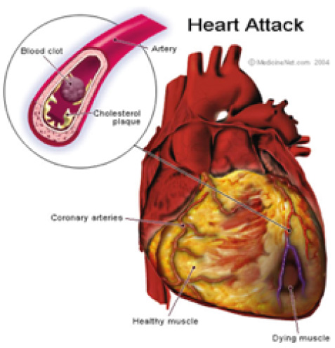
Cord Lining Stem Cells have been used to generate new heart muscle to replace the fibrous tissue from previous myocardial ischaemia to restore it to as-close-to full working pump strength as possible. 

Currently, our collaborators, Professor Sonja Schrepfer and Professor Robert Robbins from the University Heart Centre in Hamburg, Germany and Stanford University, USA, respectively are looking at immunological aspects for transplantation of Cord Lining Stem Cells for this application.

Professor Schrepfer’s team has now published two papers characterising the immunological characteristics of Cord Lining Mesenchymal Cells.

The first paper characterises the mechanisms of immunosuppression in Cord Lining Mesenchymal Cells which not only prevent them from being rejected, but also prevent other transplanted cells (like Cord Blood Stem Cells) from being rejected — thus improving their engraftment in the recipient. This paper demonstrates that Cord Lining Mesenchymal Cells appear to be superior to bone marrow mesenchymal stem cells in their immunosuppressant, and thus transplantation, qualities.

Publication: Deuse T, Stubbendorff M, Tang-Quan K, et al. Immunogenicity and immunomodulatory properties of umbilical cord lining mesenchymal cells. Cell Transplantation. 2011; 20: 655-667

The second paper compared Cord Lining Mesenchymal Cells with other mesenchymal stem cells derived from gestational tissue, namely Wharton’s Jelly, placenta and cord blood. It was demonstrated that Cord Lining Mesenchymal Cells were superior to all these other stem cells in terms of proliferation, migration, immunogenicity and immunmodulatory capabilities.

To take a quote from this publication:

“CL-MSCs showed the most promising potential for a cell-based therapy, as the cells showed low immunogenicity, but they also showed enhanced proliferative and migratory potential. Future research should concentrate on the best disease models in which CL-MSCs could be administered.”


Publication: Stubbendorff M, Deuse T, Hua XQ, et al. Immunological properties of extraembryonic human mesenchymal stromal cells derived from gestational tissue. Stem Cells Dev. 2013; (22)19: 1-9


In Professor Screpfer’s own words:

“In addition, these studies show, for the first time, that although immunomodulatory molecules HLA-G, HLA-E, and TGF-β play an important role in MSC immune evasion, intrinsic HLA expression of CL-MSCs seems to be decisive in determining the immunogenicity of MSCs. It is recommended that more research emphasis be placed on CL-MSCs and Cord Lining be the tissue of choice to be saved at birth with Cord Blood.”


Another collaborator, Associate Professor Theodore Kofidis at the National University of Singapore, Is looking at the use of multicellular angiogenic spheroids to improve Cord Lining Stem Cell engraftment in injured heart muscle.

Associate Professor Theodore Kofidis’s team has since published the work described above to show that Cord Lining Mesenchymal Cells embedded in spheroids transplanted onto the heart muscle of mice in cardiac failure reduced the symptoms of cardiac failure and improved cardiac function. This exciting work demonstrates the potential for this to be extrapolated to humans with heart failure in the future.

Publication: Lilyanna S, Martinez EC, Vu TD, et al. Cord lining-mesenchymal stem cells graft supplemented with an omental flap induces myocardial revascularization and ameliorates cardiac dysfunction in a rat model of chronic ischaemic heart failure. Tissue Eng Part A. 2013 Jun; 19(11-12): 1303-1315


The sensory receptors for hearing and balance are called neuronal hair cells, and are found in the inner ear. Damage to these hair cells from age or disease can cause hearing loss or difficulties with balance with symptoms of giddiness, nausea and vertigo. Generally, hair cells in man do not regenerate after they are lost.

Our collaborator, Professor Stefan Heller at Stanford University, has managed to differentiate Cord Lining Stem Cells into neuronal hair cells. These can be further studied to turn off the molecular ‘switch’ that prevents hair cell regeneration, or they may be directly transplanted to replace the lost cells.

her3.jpg